Researchers Establish 3D Facial Image AI Models to Predict Biological Age and Impact of Lifestyle on Aging Rate
On 7th Sep., 2020, Jing-Dong Jackie HAN’s lab from Peking University and CAS-MPG Partner Institute for Computational Biology, Shanghai Institute of Nutrition and Health of the Chinese Academy of Sciences, together with ZHOU Yong’s lab from Shanghai Jiao Tong University School of Medicine, published a research article named “3D facial image analysis to predict biological age, heterogeneity of aging rate, and impact of lifestyles” in Nature Metabolism.
Aging is the main risk factor of multiple complex diseases. People with the same chronological age may have different biological age. Currently most aging studies are based on lifespan rather than aging itself. Aging is not the same as lifespan. Aging study pursues high quality of life at old age. Systems level quantitative aging rate models, also known as aging clocks, are used for biological age calculation, and the difference between biological and chronological age is used to quantify aging rate.
In practice, due to the lack of a gold standard biological age, researchers often use chronological in place of biological age to train aging clocks and define outliers, and then support with other physical or molecular parameters. This is the common practice for existing models. For example, transcriptome aging clock based on peripheral blood mononuclear cells achieved an error of 7.8 yr (Peters et al., 2015). DNA methylation of human whole blood (Illumina 27K chip) achieved an error of 4.9 yr (Hannum et al., 2013). However, because the transcriptome or DNA methylome has to be measured in blood cells or other tissues, invasiveness and the high costs preclude their application to large-scale screenings and routine physical examinations.
Dr. Jing-Dong Jackie HAN’s group published the first 3D facial image-based age predictor in 2015 and garnered the attention from the public. This technology achieved an error of 6.2 yr (Chen et al., 2015), and compared to other aging clocks, it is non-invasive and economic, thus allowing rapid and large-scale data collection.
This newly published study extended the facial age prediction to a new AI based model by collecting 3D facial images from a cohort of ~5,000 Han Chinese, together with baseline information. The researchers established a robust non-linear age predictor that achieved an average error between chronological/perceived age and predicted age of only ±2.79/2.90 yr. Perceived age is one of the established measurements of biological age. The AI model trained on perceived age demonstrates that AI learns the human age-perceiving process, hence avoids using prediction errors as surrogates for the rate of aging. Researchers found that heterogeneity of aging rate peaks at middle age. They also profiled the transcriptomes of peripheral blood mononuclear cells using ribo-minus RNA-seq of 280 individuals and identified transcriptomic changes and cell types associated with facial aging rate.
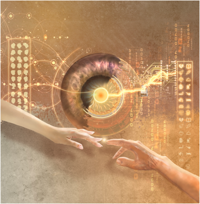
By imitating human perception of facial age, AI allows accurate determination of biological aging rate of the face, and detecting the aging interventions by lifestyles. (Image created by LI Jiaxin from Tsinghua University)
Transcriptomes, 3D facial images and lifestyle questionnaires in this study provide an unprecedented chance to find the molecular mediator by causal inference. The study found that smoking is positively related to aging rate through changes in cytokines such as SEMA6B, GRN and interferon regulatory factor (IRF8). Lifestyles such as drunkenness and preserved food consumption are also positively related to aging rate. On the contrary, yogurt, coffee, fruit, egg and bean consumptions, or meals on time are negatively related to aging rate, through increasing the level of the histone acetylation complex component ZZZ3 and repressing SEMA6B and SMAD1, etc.
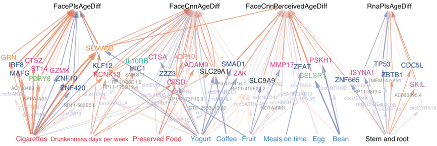
Inferred molecular mediators between lifestyles and AgeDiffs. (Image provided by Dr. Han's lab)
These relationships are deposited and visualized in the human blood gene expression-3D facial image (HuB-FI) database (http://www.picb.ac.cn/hanlab/hub-fi/).

HuB-FI database. (Image provided by Dr. HAN's lab)
This work is done by Dr. XIA Xian and Dr. CHEN Xingwei from Jing-Dong Jackie HAN’s lab as co-first authors. Corresponding authors are Dr. Jing-Dong Jackie HAN and Dr. ZHOU Yong, with help from Dr. Carlo Vittorio CANNISTRACI, Dr. ZHANG Kang, Dr. Brian KENNEDY and Dr. WANG Wei, and funding support from the National Natural Science Foundation of China, the Ministry of Science and Technology and the Shanghai Science and Technology Committee.
Media Contact:
WANG Jin (Ms.)
Shanghai Institute of Nutrition and Health,
Chinese Academy of Sciences
Email: sibssc@sibs.ac.cn